Microscopes within the BALM facility

A video of the Nikon A1R Inverted Confocal/TIRF in use by one of our PhD students.

 
The following is a selection of the microscope facilities available:

Zeiss LSM900 Confocal with Airyscan2


lsm 900

Our system is equipped with LSM 900 2 channel GaAsP URGB on Axio Observer 7 with laser lines 405, 488, 561 and 640nm, DIC, transmitted light detection on Electronically Switchable Illumination and Detection module (ESID), ZEN software including real time control electronics and ZEN software. With Definite focus for long term live cell imaging, with incubator cabinet, CO2, humidity and temperature control. Including the unique patented Airyscan technology. The innovative Airyscan detector transforms your confocal microscope into a superresolution microscope with up to 1.7x higher resolution in all three dimensions. 32 optical detection elements are arranged in an extremely light-efficient honeycomb structure, with hardly any light losses in the transition zones due to micro lenses. ZEN 3.0 system Hardware License Key Image acquisition and processing under 10 x64. User interface configurable, control of the Carl Zeiss microscope systems and components, extensive acquisition and analysis. CZI image format. Zen imaging software includes several modules including: ZEN Module Measurement-ZEN Module Multi Channel-ZEN Module Panorama-ZEN Module Manual Extended Focus-ZEN Module Image Analysis-ZEN Module Time Lapse-ZEN Module Z Stack-ZEN Module Extended Focus - ZEN Module Autofocus - ZEN Module Colocalisation - ZEN Module Spectral Unmixing (LSM only) - ZEN Module Connect Entry New with ZEN 3.0: - ZEN Module 3Dxl

Nikon A1R Inverted Confocal/TIRF

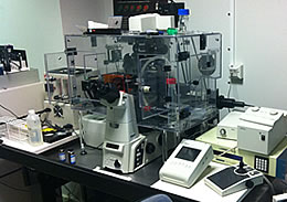
This Nikon system is a dual TIRF and laser scanning confocal (A1R) system, equipped with a Ti inverted microscope, Perfect Focus System (PFS), piezo-z, multi laser beds, a full in vivo imaging set up (T- and CO2- controlled within an incubation chamber) and an inverted motorized stage suitable for live imaging of cells (well plates or Matek dishes).

The A1R laser scanning confocal system feautures, spectral detectors, 7 laser lines (405- 457- 476- 488- 514- 546- 633nm), a resonant scanner for fast processes in live cells, live ratiometric imaging, tiled imaging optical sectioning for specimens that extend beyond the field of view (e.g. plants, invertebrates, neurons, microfluidic devices), widefield detector with DIC for simultaneous detection of confocal and transmitted light images; time-lapse imaging module and lambda imaging for spectral characterisation.

The TIRF system feautures 3 laser lines (488- 546- 647nm), an Andor EMCCD camera and 2 powerful TIRF lenses. The Nikon system is located on the 8th floor of the Biosciences building.

Nikon Eclipse E600

Nikon E600The Nikon E600 is an upright widefield/epi-fluorescence microscope equipped with an Hamamatsu CCD camera and controlled by NIS-Elements acquisition software. The system is located on the 8th floor of the Biosciences building. 


Nikon SPZ1000

nikon-spz1000This is a fluorescence stereo/dissecting microscope equipped with an Nikon DS-Fi1 camera and controlled by NIS-Elements acquisition software. The system includes and anti-vibration platform to allow minimal impact on dissection and micromanipulation and a 488nm filter. The system is located on the 8th floor of the Biosciences building.


Zeiss Elyra 7 SIM/SMLM

Elyra7

Our system is an ELYRA 7 (type LS) microscope for Photoactivated Localization Microscopy (PALM, dSTORM and PAINT) and 3D Lattice Structured Illumination Microscopy (SIM) with 4 laser lines (405nm, 488nm, 561nm and 640nm), control electronics and ZEN 3.0 SR. Together with an Axioscan frame, there is a PCO Edge camera and full incubation system and all inserts for dishes, plates chamber slides and glass slides.

The system features a Lattice laser technology (3D Lattice SIM) with optimized resolution for each wavelength for single sequential channel acquisition. Patented Lattice SIM technology is used for best depth penetration of samples in SIM (penetration up to 100 um into sample).

High speed acquisition is also possible:

  • 17 SIM image frames per second at 512 × 512 resolution and 1 ms exposure time using 15 phase images per one SIM image) and up to
  • 255 SIM image frames per second in Burst mode Lattice SIM in full resolution.

Within a ZEN processing computer, ZEN 3.0 software includes a SIM module and a SMLM module also available off line (separate workstation).

https://www.zeiss.com/microscopy/int/about-us/press-releases/2018/zeiss-elyra-7.html

Olympus Widefield/TIRF

This Olympus system is an inverted widefield/TIRF laser scanning confocal system equipped with a IX81 inverted microscope, and a full in vivo imaging set up (T- and CO2- controlled within an incubation chamber). The system feautures 2 powerful TIRF lenses, a dual view module for multicolour TIRF applications, an Hamamatsu ImageEM camera, 3 solid state lasers (491- 560- 640nm) and inverted motorized stage (suitable for live imaging of cells on well plates or Matek dishes), widefield detector with DIC for simultaneous detection of confocal and transmitted light images. The system is located on the 8th floor of the Biosciences building.

Zeiss LSM 710 Confocor 3

Zeiss-LSM710

The Zeiss LSM710 ConcoCor3 System scanner has high sensitivity and is equipped with 2 standard PMTs and 1 transmitted light T-PMT, including a tuneable AOTF and capable of imaging up to 6 channels in multitrack configuration. The main stand body is an AxioObserver inverted, equipped with a heated plate for live imaging and multiple interchangeable stage adapters to allow imaging from glass slides, chamber slides, well plates and 35mm dishes. Focus stabilisation is through software and hardware based mechanisms. The system is controlled by the Zen 2012 software capable to capture in 3D and 4D, with Lambda scanning, and spectral un-mixing capability. The Zeiss LSM710 is also integrated with a ConfoCor3 system for fluorescence correlation spectroscopy (FCS) applications.

Zeiss Cell Discoverer 7


Zeiss CD7Our Celldiscoverer 7 is configured with 7 FL channels, IR transmitted-light contrast, automated sample carrier and surface detection, 4 objectives (2x autocorr) covering 12 magnifications, applicable to 2D/3D specimen on thin and thick glass and polystyrene vessel bottom including incubation providing physiological conditions for live cell imaging. Water immersion objectives are also included. LED light source for the capture of fluorescence around example DAPI/FITC/RHODAMINE/CY5 wavelengths.

This system is an automated unit, equipped with an inverted microscope with integrated darkroom for observation of live and fixed samples. The system unit includes:

  • automatic sample container recognition, bottom thickness measurement and barcode reader
  • automatic hardware-based sample focusing and active focus stabilization
  • Reflected light illuminator FL with 7 LEDs (385 / 420 / 470 / 520 / 567 / 590 / 625 nm)
  • 5 Filter wheels for beam splitting and 7 emission filter wheel
  • Transmitted-light illuminator with IR-LED and light contrast module for phase-gradient contrast
  • Microscope CCD camera Axiocam 506 mono and CMOS camera Axiocam 702 mono
  • several inserts for plates, slides and petri dishes 35 to 60mm
  • afocal magnification changer 0.5x / 1x / 2x to provides a magnification range of 2,5x - 100x (Objectives: PApo 5x/0.35 - PApo 20x/0.7 autocorr - PApo 20x/0.95 autocorr - PApo 50x/1,2W w. Autoimmersion
  • ZEN 3.0 CD7 Software with Intellesis for automatic segmentation and thresholding. Apeer plug in for automated workflows and image analysis.

https://www.zeiss.com/microscopy/int/products/imaging-systems/celldiscoverer-7-automated-microscope-for-live-cell-imaging.html?vaURL=www.zeiss.com/microscopy/int/products/imaging-systems/celldiscoverer-7.html

.

Cryo-Fluorescence Nikon-Linkam system


Nikon E600 cryoThis is a hybrid Nikon-Linkam system equipped with a freeze plunge, Nikon E600 widefield frame with a 40x/0.6 and 100x/0.9 lenses, and an Hamamastu ORCA R2 mono camera.

This system will allow to visualize fluorescently labelled samples mounted on a EM copper grid to correlate them with TEM application (Correlative Microscopy). 

Leica DMRE

leica-dmreThe Leica DMRE is an upright widefield/epi-fluorescence microscope equipped with an Hamamatsu OrcaR2 CCD camera and controlled by RCImage acquisition software. The system is located on the 6th floor of the Biosciences building.

Leica TCS SP8

leica-tcs-sp8The Leica SP8 system is an upright laser scanning confocal system equipped with a DM6 upright microscope, AOBS spectral detector and a full in vivo imaging set up (T- and CO2- controlled within an incubation chamber). The system feautures 8 laser lines (405- 458- 476- 488- 496- 514- 561- 647nm),, a resonant scanner for fast processes in live cells, live ratiometric imaging, calcium imaging, a Hy-D detector for low phototoxicity, low signal probes and high resolution imaging; glycerol immersion objective for preservation of axial resolution when imaging in 3D, and water dipping lenses for live imaging.

Other features include: tiled imaging optical sectioning of specimens that extend beyond the field of view, e.g. plants, invertebrates, neurons, microfluidic devices; upright stand suitable for live imaging; widefield detector with DIC for simultaneous detection of confocal and transmitted light images; on-board 3D viewer and analysis software; on-board Spectral unmixing for the spectral deconvolution of multiple overlapping fluorscence; time-lapse imaging of live samples; lambda imaging for spectral characterisation.

The system is located on the 6th floor of the Biosciences building. 

In addition to training and technical assistance we also provide data analysis capabilities through our dedicated workstation

Imaging Workstations


Two PC computers are also available at no charge for imaging analisys. The software included are:

  • Fiji-ImageJ
  • NIS Elements 4.13
  • ZEN 2012
  • Leica LASX
  • ICY
  • MotionTracking