Understanding the characteristics and function of MuRF1
MuRF1 is an Ubiquitin-E3 ligase implicated in driving muscle atrophy, however there is little understood about its role in muscle protein breakdown. We aim to understand the upstream and downstream signalling pathways, drivers of expression, and potential interacting partners of MuRF1. We also aim to further this research by exploring MuRF2 and MuRF3, also implicated in muscle atrophy.
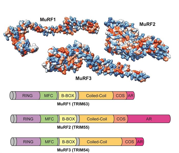
Predicted 3d structure and functional domain locations of MuRF E3 Ligases.3D structure colours denote hydrophobicity: blue=hydrophilic, red=hydrophobic. MFC = MuRF Family Conserved Domain; B-Box = B-Box Zinc Finger Domain; COS = C-terminal Subgroup One Singature; AR = Acidic Region.
Elucidating the mechanisms regulating mitophagy in skeletal muscle.
Mitochondrial turnover is crucial for maintaining cellular homeostasis. The molecular mechanisms governing skeletal muscle mitophagy (Mitochondrial degradation), particularly in response to exercise, are yet to be fully elucidated. We aim to understand the mechanisms that underlie mitophagy in skeletal muscle. This is crucial for developing therapies to combat mitochondrial dysfunction in metabolic disease.
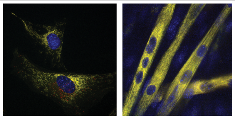
Fluorescence image showing mitophagy in action. Using GFP + mCherry tagged mitochondrial FIS1 protein, mitophagy is observable when GFP is quenched leaving mCherry alone (GFP + mCherry FIS1 construct kindly provided by Dr Ian Ganley, MRC-PPU, University of Dundee).