
Samples, Method & OCBs
Freedman & colleagues (2005) published a consensus statement dealing with CSF analysis for the diagnosis of MS. They recommended that detection of oligoclonal bands (OCB) in the CSF of patients suspected of multiple sclerosis is a “gold standard” which has excellent sensitivity (>95%) and specificity. This section describes in details some fact of OCBs with sample requirements, methodology and examples of the main patterns seen with interpretation are emphasised.
Facts about oligoclonal bands
- Two or more oligoclonal bands (OCB) in the CSF suggests intrathecal IgG synthesis.
- OCBs are indicative of immune response in the CNS and diagnosis of multiple sclerosis (MS) in patients with clinical suspicion.
- There is no correlation between OCBs in CSF and demyelinating process
- OCB can be present even when the CSF IgG level is normal
- An established oligoclonal response may exist throughout life (except in CNS infections)
- OCB also found in other cases thus OCBs in isolation are meaningless.
- Isoelectric focusing is most sensitive method for detection of the OCB.
- Patterns 1-5, described in this section, are typically common (but real examples). The important patterns in MS are those that demonstrate "intrathecal IgG synthesis" (pattern 2 and 3).
Sample requirement
For the detection of oligoclonal bands, paired serum and CSF samples are required. Blood must be taken at the same time as the lumbar puncture (LP), if not then within two to three weeks of the LP. Results cannot be relied upon beyond this time limit (half life of serum IgG is about 23 days).

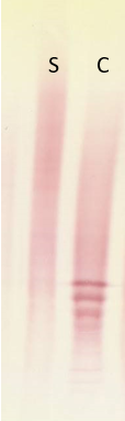
This example demonstrates the effect of unpaired samples on oligoclonal bands.
Serum was taken six weeks after the LP and contains extra bands which are not reflected in the CSF. The absence of bands in the serum corresponding to those present in the CSF might lead to incorrect interpretation of intrathecal IgG synthesis (pattern 2 and/or 3). Due to the time delay the relevant IgG may have been cleared from the serum. Consequently, these results could not be relied upon without another LP.
Isoelectric focusing method
Isoelectric focusing method
Summary
GEL SOLUTION: 0.3g agarose, 3.6g sorbitol, 27ml of 10% glycerol and microwave to dissolve.
CASTING GEL: AT 65°C add 2ml (pH 3-10) & 0.5ml (pH 8-10.5) pharmalyte to molten agarose (65-70°C), mix and cast at 65°C.
HYSTERESIS: Keep the casted gel at 4°C in a moist chamber for at least 30 min.
SAMPLE PREPARATION: Serum diluted at 1:400 & 5µl loaded. Vol. (µl) of CSF loaded = (2.5/total protein).
RUNNING THE GEL: Blot the gel with nitrocellulose membrane (NCM). Position the electrode wicks at 7cm apart (1M NaOH (-ve), 0.05M H2SO4 (+ve)) on the gel and then the application strip 2cm from +ve electrode. Remove application strip after 20mins. The gel is electrophoresed at 1250 volt/hour.
BLOTTING: Pre-blot gel for 10 sec with NCM and discard it. Blot with another NCM piece for 30min.
BLOCKING: Dry the blot. Incubate in 2% marvel/saline for 30minutes and quick rinse in tap water.
PRIMARY ANTIBODY: Anti-human IgG (gamma chain) peroxidase, ~1:200 dilution in 0.2% marvel/saline and incubate for 60mins.
WASH/TAP WATER/SALINE: Rinses in tap water followed by 5 min in saline.
DEVELOPMENT OF THE BLOT: To 50mL of 50mM sodium acetate (pH 5.1), add 20mg tablet of A.E.C. dissolved in 2.5ml D.M.F/methanol and 30µl of H2O2, (20-30mins).
WASH/TAP WATER: Rinses in running tap water, dry and store in dark.For Interpretation of oligoclonal bands. See real examples of most commonly recognised patterns of OCB elsewhere on this site.
Patterns in OCBs

Pattern 1: No oligoclonal bands in CSF or Serum. No intrathecal IgG synthesis.

Pattern 2: Oligoclonal bands present in CSF only. Intrathecal IgG synthesis as seen in MS.

Pattern 3: Identical bands in both serum and CSF with extra bands in CSF. Demonstrates both intrathecal and systemic IgG synthesis. This is also seen in MS.

Pattern 4: Bands in serum mirror those in CSF. This suggests systemic IgG synthesis.

Pattern 5: These abnormal "ladder" type identical bands seen both in the CSF and serum are usually monoclonal proteins, suggesting peripheral IgG synthesis.
KEY: S = Serum, C = CSF. The blot number also represents both the pattern and the anode side.
OCBs in other conditions
| Conditions | Pattern 2 | Pattern 3 | Pattern 4 |
|---|---|---|---|
| . | .jpg?quality=80&height=250&width=100) |
.jpg?quality=80&height=250&width=100) |
.jpg?quality=80&height=250&width=100) |
| Infection | Yes (CNS) | Yes | Yes (systemic) |
| Inflammation | Yes (CNS) | Yes | Yes (systemic) |
| Paraneoplastic | Yes | Yes | Yes |
| Neoplastic | Rare | Rare | Rare |
| GBS | No | No | No |
| Other neuropathies | No | No | Yes |
| Multiple Sclerosis | Yes | Yes | No |
| Vascular disease | No | No | Rare |
| Degenerative disease | No | No | Rare |
GBS = Guillain-Barré syndrome, S = serum, C = CSF
CSF Proteins
CSF Paraprotein
CSF Paraprotein
Evidence of intrathecal paraprotein suggestive of CNS lymphoma / CNS plasma cell dyscrasia.
NOTE: Specimen mix up was eliminated by repeating the test with another blood sample from the patient.
Tau protein (β transferrin or asialo-transferrin)
Tau protein (β transferrin or asialo-transferrin)
Tau protein (ß transferrin or asialo-transferrin) is present in cerebrospinal fluid (CSF) and is used as a marker for the identification of CSF. This is particularly useful in cases where CSF leakage occurs for various reasons such as for example head trauma, congenital malformation, tumour or surgical procedure. In addition, up to 40% of CSF leakage can be spontaneous. Leakage can occur from the nose, the ear, the eyes or from the head/neck wound after an injury.
Untreated poses serious risk of infection.
An example of positive Tau protein.
+ (anode)
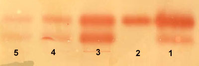
This blot shows positive identification of CSF in nasal fluid. In tracks 3, 4, and 5 there are two bands, in the same position as the control CSF (track 1).
An example of negative Tau protein.
+(anode)
.jpg)
This nasal fluid does not contain CSF as it resembles the pattern seen with serum control (track 2).
Key: 1 = control CSF, 2 = control serum, 3 = neat nasal fluid, 4 = 1/5 diluted nasal fluid and 5 = 1/10 diluted nasal fluid