
Advanced Microscopes and Equipment
For further information, including access and fees, please visit our COMPARE Advanced Imaging Facility site or contact compare@contacts.bham.ac.uk
Cairn's Quad TIRF Microscope
Cairn's Quad TIRF Microscope
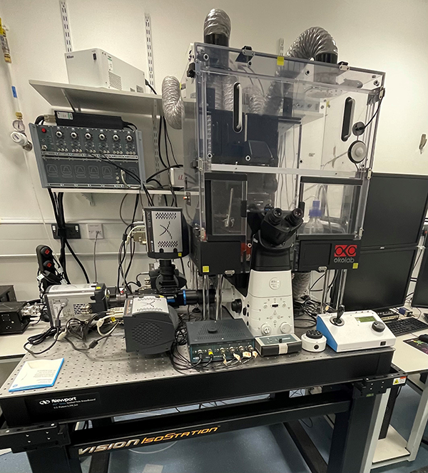
This custom 4-camera TIRF is capable of simultaneously imaging up to four different molecules (labelled with different colours) with single-molecule sensitivity, permitting analysis of movement of individual molecules as they diffuse and interact in living cells with high spatiotemporal resolution. This provides quantitative information on complex populations of molecules, including their nanoscale arrangement, diffusion and interactions.
Microscope Details
- Sample size: single cell
- Main application: protein interactions/SMLM
- Modalities: SMLM, TIRF, dSTORM (2D and 3D), PALM (2D and 3D), FRAP, FRET
- Spatial Resolution: ~20 nm lateral, ~50 nm axial
- Temporal resolution: fast (~50 fps)
- Environmental control: Tempterature and humidity
- Excitation Lasers: 405, 445, 488, 514, 561 and 637 nm
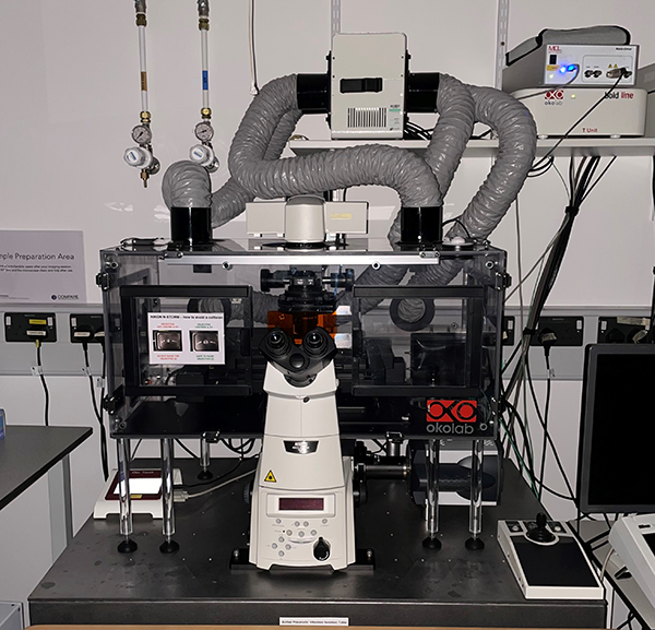
Nikon's N-SIM S Microscope
Nikon's N-SIM S Microscope

SIM (Structured Illumination Microscopy) is a form of super resolution microscopy that allows living and fixed fluorescent samples to be observed at approximately twice the resolution of a standard fluorescence microscope. Imaging at this higher resolution can reveal structural sub-cellular organisation which would otherwise be masked on a standard microscope.
Microscope Details
- Sample size: single cells, organoids
- Main application: Structured Illumination Microscopy (SIM)
- Modalities: 2D and 3D SIM, TIRF SIM
- Spatial resolution: ~115 nm lateral, ~270 nm axial
- Temporal resolution: medium (~15 fps)
- Environmental control: Temperature, humidity and CO2
- Excitation Lasers: 405, 488, 560, 640 nm
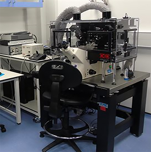
Nikon N-STORM Microscope
Nikon N-STORM Microscope
Stochastic Optical Reconstruction Microscopy (STORM) and Photoactivated Localization Microscopy (PALM) are based on recurrently determining the position of single fluorescence emitters in live or fixed samples. The repeated localizations of individual molecules is then used to reconstruct super-resolved images. Applications include studying the nanodomains organization of signalling complexes or small subcellular structures like mitochondria or the cytoskeleton.
Microscope Details
- Sample size: Single cells, tissue slices, organoids
- Main application: Stochastic Optical Reconstruction Microscopy (STORM)
- Modalities: dSTORM (2D and 3D), PALM, TIRF
- Spatial Resolution: ~20 nm lateral, ~50 nm axial
- Temporal resolution: medium (20 fps)
- Environmental control: Temperature, humidity and CO2
- Excitation Lasers: 405, 488, 560, 640 nm
Ultrafast FRET/calcium imaging Microscope
Ultrafast FRET/calcium imaging Microscope

This Ultrafast FRET/Calcium imaging microscope is equipped with a fast perfusion system for rapid stimulation and an image splitter for synchronous dual-channel imaging. Combined with a series of FRET sensors and indicators, this system allows monitoring of the kinetics of cell signaling events such as receptor activation or the production of second messengers such as calcium or cyclic AMP in living cells with unrivalled temporal resolution.
Microscope Details
- Sample size: single-cells, organoids, acute tissue slices
- Main application: FRET and ratiometric imaging
- Modalities: FRET, Ca2+, dual-channel ratiometric imaging
- Environmental control: Temperature, humidity and CO2
- Illumination: 8-channel LED illuminator
- Resolution: 200-300 nm, up to 300 ns.
3i's Lattice Light Sheet Microscope
3i's Lattice Light Sheet Microscope
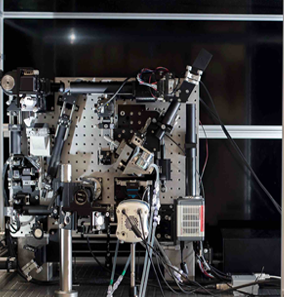
The lattice light-sheet microscope is the leading fluorescence microscope for imaging dynamic cellular and sub-cellular processes in single cells. Using this microscope you will be able to gain precise information on protein and organelle dynamics, with the highest and most accurate time resolution currently available in a fluorescent imaging system.
Microscope Details
- Sample size: single cell
- Main application: fast subcellular dynamics in live cells, with low phototoxicity
- Modalities: 2D optical lattice with ultra thin sheet (<600 nm)
- Spatial Resolution: ~230 nm lateral, ~70 nm axial
- Temporal resolution: ~300 z-slices/sec
- Environmental control: Temperature only
- Excitation Lasers: Bessel beam light sheet illumination in 488, 561, 640 nm
3i's Marianas Light Sheet (diSPIM) Microscope
3i's Marianas Light Sheet (diSPIM) Microscope
Selective plane illumination microscopy (SPIM) on the Marianas Light Sheet uses a thin sheet of light to illuminate only the plane of interest, reducing phototoxicity by drastically cutting total light dose and allowing for prolonged specimen imaging.
The system captures two alternating views of a sample which are then merged to generate an even 3D image without distortion, and is suitable for imaging cellular and subcellular structures in larger live samples (tissue sections, spheroids/organoids, small organisms).
Microscope Details
- Sample size: cells and small tissue, embryos, organoids, etc.
- Main application: rapid 3D imaging with isotropic resolution and low phototoxicity
- Modalities: Light sheet selective plane illumination microscopy (SPIM)
- Spatial Resolution: isotropic 330 nm x, y, z
- Environmental control: Temperature, humidity and CO2
- Imaging media: Aqueous media only
- Excitation Lasers: 488, 561 and 640 nm
Miltenyi's Ultramicroscopell Microscope
Miltenyi's Ultramicroscopell Microscope
The UltramicroscopeII is a fluorescent microscope used for 3D imaging of optically-cleared samples such as whole mouse organs and small tissue samples (essentially 3-D histology).
It is best suited for visualizing whole biological systems with cellular resolution.
The system uses six light sheets in a dual-sided arrangement for homogeneous illumination, with the detection objective perpendicular to laser illumination.
Microscope Details
- Sample size: whole organs, large tissue, embryos, etc (< 1cm3)
- Main application: 3D imaging of whole biological systems
- Modalities: uni and bi-directional imaging with variable sheet width
- Spatial Resolution: cellular
- Environmental control: N/A
- Excitation Lasers: 488, 561, 640 and 780 nm
ONI Nanoimager Microscope
ONI Nanoimager Microscope

The Nanoimager is a multivalent microscope for super-resolution microscopy, covering dSTORM, PALM, single-particle tracking, TIRF, HILO and smFRET.
Like other super-resolution systems, it allows the study of high-level molecular organisation on fixed samples.
Microscope Details
- Single-molecule sensitivity
- Sample size: Single cell
- Main application: single-molecule localization microscopy (SMLM)
- Modalities: dSTORM (2D and 3D), PALM, SPT, TIRF, FRET
- Spatial Resolution: ~20 nm lateral, ~50 nm axial
- Temporal resolution: medium (20 fps)
- Environmental control: Temperature, humidity and CO2
- Excitation Lasers: 405nm, 488nm, 561nm, 640nm
LUMICK’s C-TRAP
LUMICK’s C-TRAP

The C-Trap combines optical tweezers, confocal fluorescence microscopy and microfluidics to measure molecular interactions, forces and dynamics at the single-molecule level. It enables simultaneous manipulation and fluorescence imaging of biomolecules, including DNA, proteins and molecular complexes.
Microscope Details
- Sample type: Purified biomolecules, DNA–protein complexes, molecular assemblies and cell-derived extracts
- Main application: Single-molecule force spectroscopy and fluorescence imaging
- Modalities: Optical trapping, confocal fluorescence, force detection and automated microfluidics
- Optical traps: Four dynamic optical traps
- Force detection: Dual-axis force detection in x and y
- Fluorescence channels: 488, 561 and 638 nm
- Positioning and tracking: Active force calibration, trap stabilisation, piezo-based trap tracking and 3D nano stage
- Environmental control: Temperature control
- Microfluidics: Automated multichannel microfluidic control
- Typical applications: DNA–protein interactions, molecular motor activity, protein folding, receptor–ligand interactions and mechanobiology

Sample Preparation Lab
In support of all our advanced imaging systems, the facility also provides a dedicated space for sample preparation.
The sample preparation lab is available to all our users, and features common wet lab and tissue culture equipment, including TC incubators, laminar flow hood, and an EVOS FL microscope to allow for basic sample checks of cell density and fluorescence.