Pulp Biology and Regenerative Endodontics
Staff: Cooper, Scheven, Shelton, Tomson
Our Pulp Biology and Regenerative Endodontics research programme aims to better understand the relationship between the tissue injury events which occur during disease and regenerative processes, focusing on stem / progenitor cells involved in the regenerative events along with matrix-mediated cellular signalling processes. The selection and isolation of stem cells with dentinogenic potentiality is now providing a tangible approach for clinical translation to develop new regenerative therapies. This work is further facilitated through our studies on novel hydrogel technologies which may be used to enable dentinogenic repair.
The mechanistic basis of dental materials such as calcium hydroxide in pulp capping regenerative therapies has long been studied and our recent work has now demonstrated that this restorative material along with others such as Mineral Trioxide Aggregate (MTA) and Biodentine, can locally release matrix-bound growth factors which beneficially modulate pulp cell responses. Indeed our work is making significant contributions at the cutting-edge of regenerative endodontic research by characterising novel growth factors in this process and determining their modes of action in fundamental tissue repair-associated processes such as cell proliferation, differentiation, angiogenesis, chemotaxis and mineralisation. We are also making significant in-roads into our understanding of the interactions between the inflammatory and tertiary dentinogenic responses.
Research has identified several molecules that have pleiotropic effects and that the doses and temporal expression of these factors along with interaction with matrix molecules may be critical to their cellular signalling of inflammatory/immune or regenerative events in the diseased pulp.
Craniofacial Tissue Regeneration and Engineering
Staff: Cooper, Shelton, Scheven
Our research in this area is aimed at obtaining a better understanding of the therapeutic potential of a range of stem cell-types and their role in craniofacial tissue regeneration. We are working with stem cells derived from bone marrow, adipose, dental pulp and periodontal ligament tissues and are characterising their roles and responses in 2D and 3D standard culture and biomimetic environments. Our aims include the identification and generation of conditions that can be used to better understand the role of these cell types in tissue repair.
Our studies have compared the effect of cell sorting, cryopreservation and differentiation approaches on different stem cell types. Furthermore we are characterising the role of the secretomes and exosomes on osteo-genic/-clastogenic, angiogenic and immune cell responses. Our findings have already highlighted that different stem cells cultured under different conditions in vitro may offer different therapeutic potential via the molecules they release. These findings may have significant therapeutic potential for tissue repair in dental diseases such as caries and periodontitis as well as in craniofacial bone regeneration following disease or trauma.
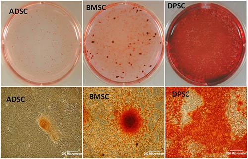 (Above) Osteogenic differentiation in cultures of adipose-derived stem cells (ADSC), bone marrow mesenchymal stem cells (BMSC) and dental pulp stem cells (DPSC) showing various patterns and degrees of mineralisation.
Neurogenesis and neuronal repair
Staff: Scheven, Mead (PhD student 2012-2015)
Colaborators: Logan, Leadbeater
Our research group is evaluating the role and application of dental mesenchymal stem cells for neural repair. Our multi-disciplinary and collaborative research uses specialised models to study the neurogenic cell differentiation potential of dental and mesenchymal stem cells as well as the role and effects of stem cell-derived trophic factors (secretome and exosomes) in neural injury and disease. Current focus is on the therapeutic use of DPSC in retinal & optic nerve injury following (head) trauma or neurodegenerative disease such as glaucoma. We have obtained promising evidence that DPSC represent a potentially advantageous paracrine-mediated cell therapy for neuroprotection of retinal neuronal cells (retinal ganglion cells) and retinal nerve axon regeneration. Research is aiming to elucidate the precise trophic mechanisms of action by the stem cells and the development of a clinically suitable & translatable stem cell-based therapy.
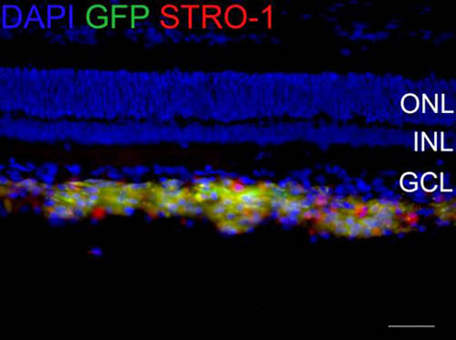
(Above) Localisation of GFP-transfected DPSC within the vitreous body of the eye following cell transplantation
Theraputic Ultrasound
Staff: Walmsley, Scheven, Cooper, Shelton, Patel
External colaborators: Prof SR Ghorayeb (Hofstra University, NY, US)
Ultrasound has various medical applications. Our research group is exploring the use of ultrasound as a non-invasive physical therapy for dental tissue healing and repair. The tooth is a hard mineralised structure with a living soft pulpal tissue core capable to respond to outside stimuli including physical forces. We study how ultrasound penetrates and travels in dental tissues and how it affects the reparative activities of the living cells within the core of the teeth. Our research has demonstrated that ultrasound in the low frequency range, generally used in dentistry for dental scaling, is able to stimulate both odontoblasts and dental pulp cells which are involved in dentine formation and repair. The research represents a true multidisciplinary collaboration involving experts from physics, engineering, biology, biomaterials and dentistry. Mathematical simulation models are being developed to analyse low frequency & low intensity ultrasound transmission within dental tissues which will be compared with ultrasound-induced bio-effects in different experimental tooth models. Our studies also investigate whether ultrasound is able to activate and stimulate stem cells to support healing of dental and bone tissues. The outcome of our studies is to exploit the development of a new ultrasound and non-invasive therapeutic devices that can be used in the clinic for dental, periodontal and craniofacial repair, thereby promoting oral health and ultimately human health.
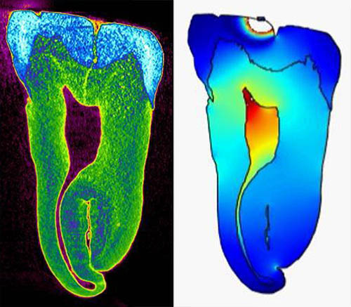
(Above) CT scan of a tooth (left panel), and pressure and intensity plots of (45 kHz, 31 kPa) ultrasonic wave throughout the tooth including central pulp chamber (right panel).
(From: Ghorayeb, Patel, Walmsley, Scheven, J Ther Ultrasound 1:12, 2013)