Dr Steve Mayhew uses novel imaging techniques to navigate the outer limits of the brain. Ros Dodd learns more.
As a physics undergraduate back in 2002, Steve Mayhew’s ambition was to become a space scientist. But as time went on, he switched to a subject even more shrouded in mystery – the human brain.
During postgraduate studies at the University of Oxford, Steve decided biological systems were more interesting and he became especially fascinated by the enormous challenge of understanding how the brain functions.
‘I did a degree in physics at the University of Bath and initially I wanted to be a space scientist,’ he recalls. ‘But during a placement year at Oxford, I became more interested in living things and I started to focus on neuroscience. A lot of interesting things happen in our skulls.’
And so began the research work that Steve, now Dr Mayhew, is further developing as a Birmingham Fellow within the University’s School of Psychology – applying physical techniques to problems in neuroscience.
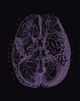
This focuses on neuroimaging; using simultaneous recording of electroencephlography (EEG) during functional magnetic resonance imaging (FMRI).
This mapping of the brain can help scientists like Steve understand more about how the brain works – especially when it’s affected by ageing or disease such as dementia, epilepsy and stroke.
‘I found the idea of medical imaging fascinating – to use physical properties to gain an insight into tissue structure and follow the function of the processes. Once I got into brain imaging, I never looked back.’
Although based in the School of Psychology, which houses the state-of-the-art Birmingham University Imaging Centre, Steve doesn’t consider himself a psychologist. ‘I’m not sure what I am – a neuroscientist, I suppose,’ he smiles.
‘My work focuses on understanding the techniques we use for imaging and what the measurements we make can tell us about brain signals. My primary interest is in using functional MRI techniques (FMRI), which take advantage of the properties of the body tissue and can create a whole array of different images of it, depending on what exactly you’re interested in.
‘Very simply, you can use MRI to obtain structural images, like a high-resolution X-ray; but we can also use MRI to measure brain function. As blood flows through your brain, nutrients like glucose and oxygen are extracted from it as a result of the neuronal activity in a given area. This enables you to map that activity with an FMRI scan. So FMRI is sensitive to the magnetic properties of the blood, which depends upon the oxygen content, and this can tell you a good deal about where the brain’s different functions can be located.’
Although we know quite a lot about the basic anatomy and functions of the brain already – that it has a right and left hemisphere, for example, with the right hemisphere controlling the left side of the body and vice versa – there is much still to be discovered.
But scientists have made much headway in the past 15 years or so, since neuroimaging became possible.
‘That’s been long enough to map pretty much all the obvious functions of the brain you can think of,’ explains 32-year-old Steve. ‘At the moment, a lot of the easy things to study with brain mapping have been done. So what we’re trying to do is to push the technique a bit further – and that’s where things are going to get complicated.’
Because FMRI measurements are based upon blood flow, it is only an indirect measurement of brain function. So Steve and his team combine FMRI with EEG, a technique that has been around for about 100 years. This involves attaching an array of electrodes to the scalp and recording the voltage.
‘One of the things we specialise in is recording EEG simultaneously with FMRI to enable you to record both aspects of brain function. EEG measures brain activity directly and is simple, relatively cheap and easy to apply. It’s been used clinically for a long time; for example, to help diagnose epilepsy.’
However, EEG has its own drawbacks. While it can record neuronal activity with high temporal precision, its ability to specifically localise the source of that activity is limited.
‘There are strengths and weaknesses with all these techniques, even when they’re combined: they don’t give us an optimal measurement. But the advantage of combining FMRI and EEG is that you get the best out of both techniques and they are very complementary measurements. We’re trying to maximise this and get the most from them.’
Although FMRI is ‘brilliant’ in terms of producing a high-resolution map of where things happen in the brain, it’s quite slow because it relies on blood flow and blood flow can’t keep up with the brain’s workings. But EEG, because it directly measures neuronal activity, is instantaneous.
‘So the great advantage is that by combining the two things, you not only get more temporal information to go with the FMRI signal, you can compensate for FMRI not being a direct measurement of brain activity,’ explains Steve. ‘This is useful, because although we understand the underlying biology of the healthy brain, in disease and in ageing, the biology changes. That means the FMRI signal can change – but that might not reflect a change in neurological activity; it could simply be a blood flow change. If you have recorded EEG measurements as well, then you have a way of controlling that.’
A large body of research has been done on Alzheimer’s disease, which is becoming more prevalent as people live longer.
‘A lot of research is done on Alzheimer’s,’ says Steve. ‘We have an increasingly ageing population and a lot of people are struggling with these kinds of diseases.’
Scientists still don’t really understand what happens when people’s memories start to break down – why they can no longer retain facts as they used to do. Is it because of restricted blood flow to the brain, or is it due to changes in neuronal activity?
‘I have done some work in elderly people to try to develop a technique where we can estimate this better and we can get better measurements, better quantification in what the FMRI image represents. I’m also starting some work with computer models to try to test theoretically how different combinations of biological factors might give rise to variations in the imaging signals that we record – the idea being that if you can simulate data, you can test what happens if you change a parameter in the system. You can make predictions from that and then test them in new experiments.’
Because the brain isn’t hard-wired at birth – as used to be thought the case – and has plasticity, it’s possible for it to ‘relearn’ functions it has ‘forgotten’ by creating new circuits and pathways.
FMRI allows scientists to track the brain’s function. So, for example, if you scanned a patient immediately after they’d had a stroke and couldn’t move their right arm, you would be able to see which part of the brain was not working properly. If you continued to scan them and they started to regain use of their arm, the imaging would show which part of the brain was responsible.
But, stresses Steve, there is still a lot scientists don’t understand about even basic brain function. For instance, if you get someone to tap their fingers on a desk and look at the sensory impact on the brain, the brain’s response is slightly different every time. The same is true when mild pain stimuli are applied to someone’s leg, when beeps are played via headphones or lights flashed into someone’s eyes.
‘There seems to be no such thing as a consistent brain response and we don’t really know why that is. Why is there so much variation? Why does it happen and is it meaningful that it does? These are some of the questions that my research is trying to address.’
Steve and his team are also hoping to set up a project with Birmingham City University in which patients with chronic pain are given cognitive behavioural therapy (CBT) as a way of easing their suffering.
‘The project will focus on scanning the patients’ brains to see if there’s anything in the natural patterns of their activity to predict whether CBT is likely to help them. This would be useful because if it could be shown CBT wasn’t likely to help, you’d save money on putting them through a ten-week course and could get doctors to focus on more appropriate therapy.
‘The same with epilepsy: some types of epilepsy are easy to diagnose, but a lot aren’t. But these techniques might be able to help diagnose the trickier ones. So we’re hoping this research will prove useful in both clinical diagnosis and treatment.’