Ultrafast Microscopy

Lightwave-driven imaging at the scale of atoms
Quanmin Guo, Tom Siday
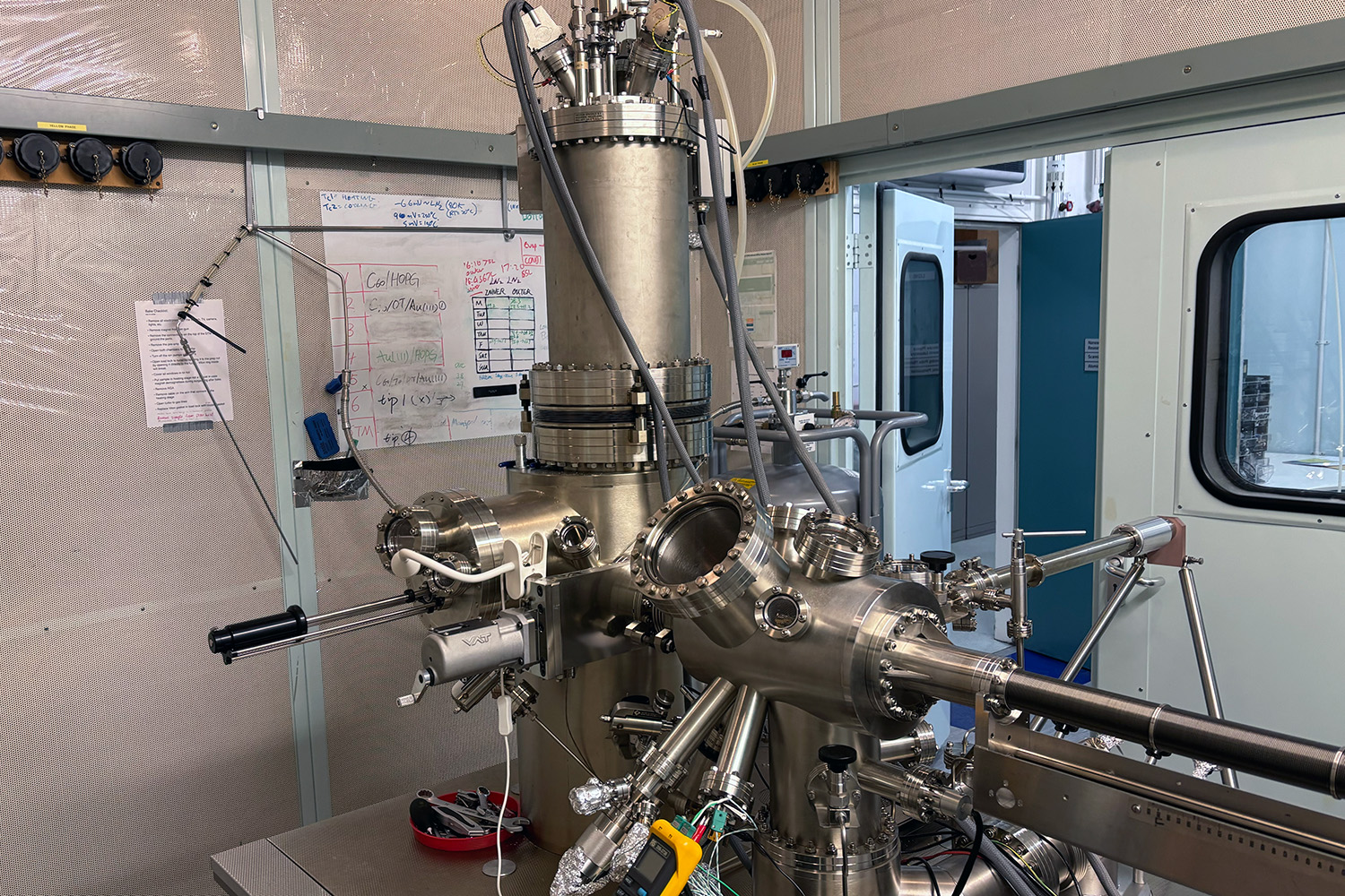
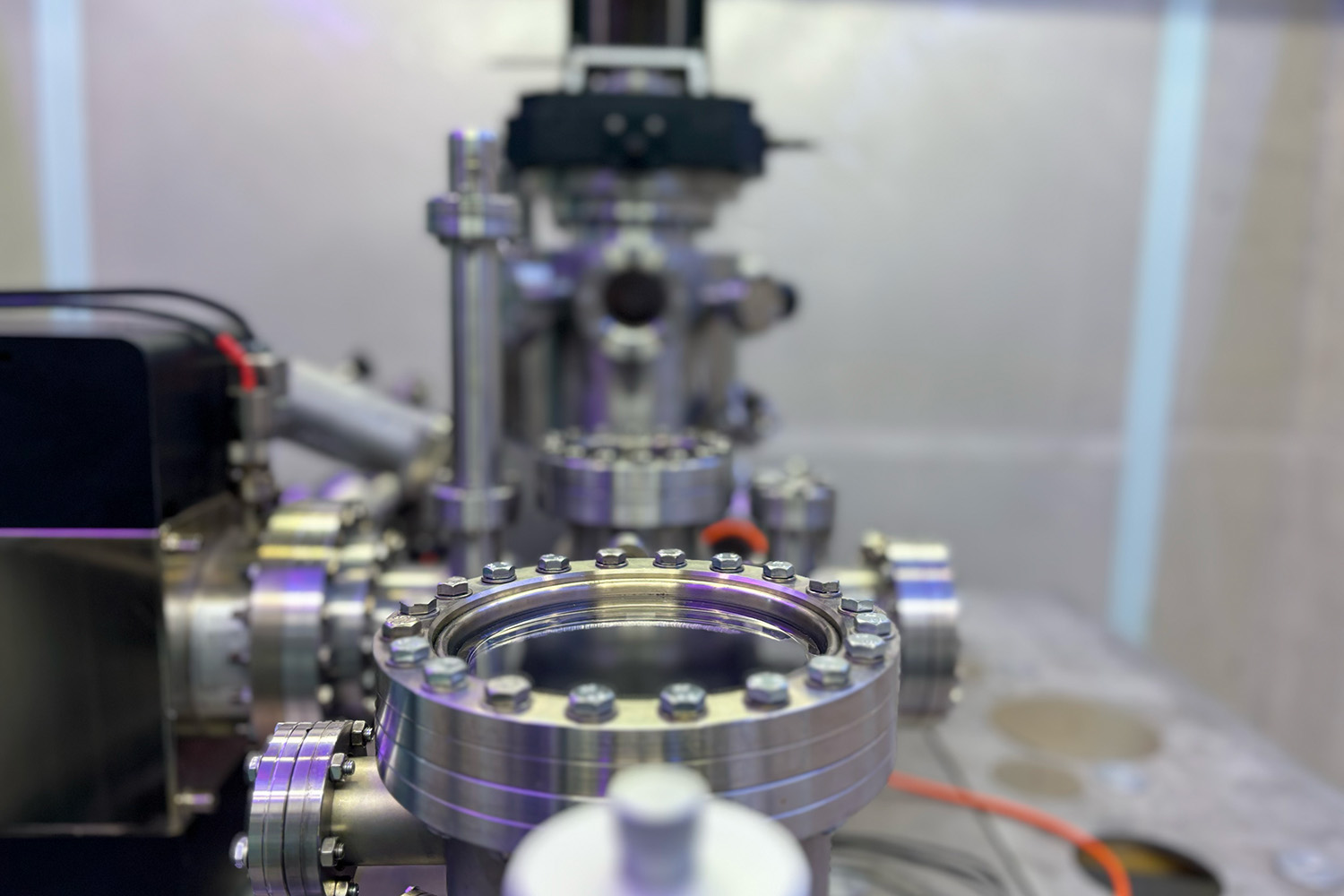
By exploiting the evanescent field enhancement at the apex of sharp metal tips we can create intense, localised light–matter interactions at the nanoscale. Combined with techniques like atomic force microscopy, this approach forms the foundation of an ultrafast nanoscope. To push beyond the nanoscale, we can harness strong nonlinear effects - like the quantum tunnelling of electrons - to access atomic resolution in both space and time. This opens a direct window onto atomic-scale dynamics with femtosecond precision.

This research is supported by multiple UHV microscopes: a low-temperature (base 4K) and variable temperature (base 25K) STM (Scienta Omicron), alongside a bespoke ultrafast UHV nanoscope (developed in collaboration with University of Oxford).

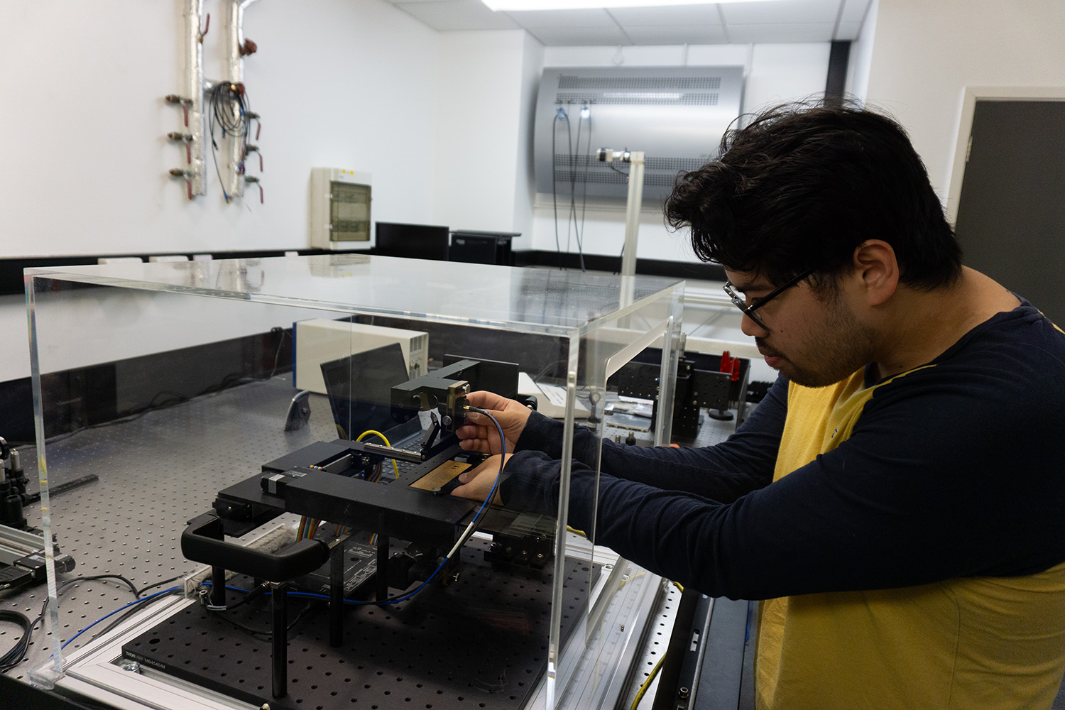
Subcycle microspectroscopy
Andre Kaplan, Miguel Navarro-Cía, Tom Siday
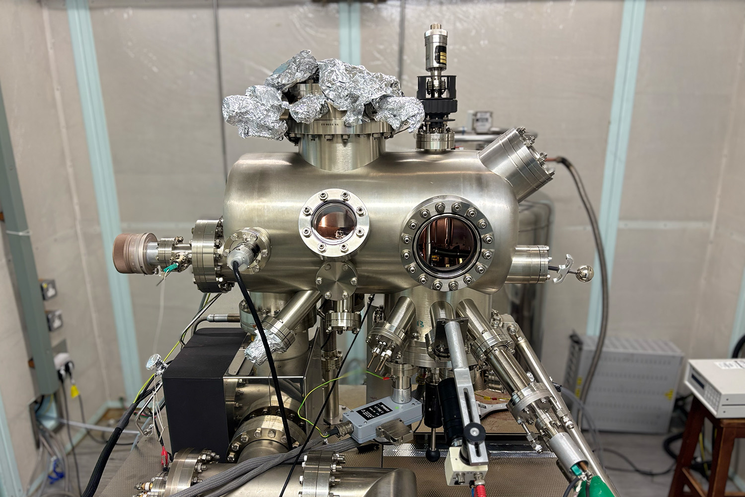
Nanoscopy directly can address light-matter interactions at their most fundamental level - yet the emergent properties of quantum materials often appear over slightly larger length scales (~1-100 µm) – especially when the energy scale of collective excitations is in the terahertz (1012 Hz) range. To access these length scales, a small aperture can be used instead of a tip to efficiently collect evanescent near fields. Doing so, we can directly address a broad range of quasiparticles - from phonon polaritons to magnons - inaccessible to conventional far-field spectroscopic techniques and providing unique insight into the nonequilibrium dynamics of the quantum world.

This research is supported by multiple terahertz near-field microscopes: one based on commercial (sub)systems (Toptica and Protemics), and a bespoke ultrafast near-field aperture microscope developed in conjunction with University College London.

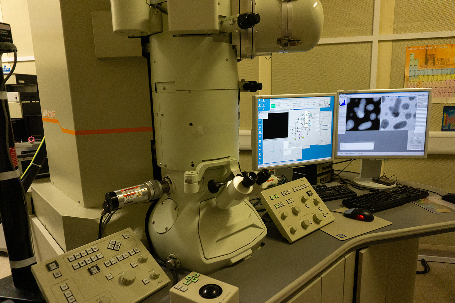
Transmission electron microscopy
Wolfgang Theis
Transmission electron microscopy is a powerful method to study nanoscale systems. It provides access to both the atomic structure and chemical composition of the studied samples.
This research is enabled by a 200kV JEOL2100 transmission electron probe microscope, with probe aberration corrector. This achieves atomic scale resolution of 0.8 Angstroem in annular darkfield scanning transmission mode.
Key experimental techniques
Key experimental techniques