
Intravital Microscopy Suite
The Intravital Microscopy Suite is an imaging facility dedicated to the delivery of high-quality and high-impact intravital microscopy. The facility has a wide range of equipment, including widefield, confocal, and multiphoton microscopes. Additional tools, such as laser ablation systems, allow us to probe vascular events such as thrombosis in fine detail. We also have tools for measurement of vascular perfusion, such as full-field laser speckle and laser doppler. In addition, we are able to support researchers with HO PIL and PPL licencing and experimental support.
The academic lead and Director of the facility is Dr Neena Kalia. Dr Dean Kavanagh is the dedicated manager and deals with the daily running of the facility and training of users in all aspects of intravital research. Between them, they have over 35 years experience in intravital microscopy.
Why intravital microscopy
Why intravital microscopy
Intravital microscopy, at its most basic definition, is the use of powerful imaging techniques to visualise events taking place in biological tissues in real-time and in vivo. These might include imaging the various steps of the leukocyte adhesion cascade, the development of platelet aggregates and thrombi, changes in vessel leakiness, flow, functional capillary density or vessel tone.
Through the use of fluorescent stains and genetically engineered animals, we’re able to image and monitor a range of cellular events dynamically in the intact physiological environment. This ability to label a number of cellular components and visualise them simultaneously, makes intravital imaging techniques useful for answering a range of biological research questions, particularly in the fields of thrombo-inflammation, tumour biology, and stem cell trafficking.
Moreover, we can utilise intravital imaging to look at general architectural changes in tissues and also non-vascular structures in vivo e.g. fibrillar collagen or muscle fibrils using second harmonic generation microscopy.
Why work with us
Why work with us
Our aim is to make intravital microscopy accessible to researchers by providing support at all steps of the process. We can provide help with licencing, whether they be new applications or amendments to existing licences. We can provide experimental advice; assisting in the choice of model and method that is best suited for your study.
We can provide training for most procedures in-house and will support students and staff to become competent in the required techniques. If you wish to bring external techniques to Birmingham, we can help you achieve this – please speak to us.
Finally, we are always available to help advise on how best to analyse and present your data for publication and presentation.
Who do we work with
Who do we work with
We currently work with research groups across the University from a wide range of departments/schools and across a variety of colleges.
We welcome the opportunity to work with anyone who has an interest in intravital microscopy or in vivo imaging.
Resources and services
Resources and services
We have five microscopes in the facility which are available for use by researchers. We have one upright widefield, two upright confocals, one inverted confocal and one multiphoton microscope.
All of our confocal microscopes are fitted with Nipkow spinning disks (Yokogawa) and are designed for high-speed live imaging. They all have fast-wavelength switchers which allow for near simultaneous imaging of multiple fluorophores in the same experiment. The confocal heads have both high-signal and high-speed filters, allowing users to choose between modalities based on their experimental needs. In addition, our upright confocal microscopes have built-in laser ablation systems that allow for the creation of targeted vascular damage and these are particularly useful in assays of thrombus formation and platelet activity.
Our multiphoton is an Olympus FVMPE-RS in a dual laser line setup. The dual laser setup allows for simultaneous stimulation of fluorophores on two laser lines, and with appropriate filter/fluorophore selection, allows for the imaging of up to four colours simultaneously. The multiphoton also has photoactivation modules, allowing for optogenetics/photoswitching studies to be performed in conjunction with intravital imaging. We have filters for second harmonic imaging, allowing the label-free visualisation of collagen and other tissue structures.
Current research
Current research
We are currently engaged in a number of projects, which cover a variety of cardiovascular, cancer, immunology and inflammation research. We have extensive experience in imaging a variety of healthy and diseased tissues and organs including the cremaster muscle, liver, kidneys, spleen, skull bone marrow, pancreas, heart etc. We also work with researchers on deep tissue multiphoton imaging of ex vivo tissues and organs. An example of our active projects include:
- Improving cell-based cancer therapeutics in the pancreas.
- Impact of co-morbidities on the coronary microcirculation.
- Alterations in large vessel thrombus formation in response to drugs and potential therapeutic compounds.
- Mechanisms governing inflammatory and immune cell recruitment in the liver, heart and kidney models of disease.
- Monitoring the dynamics of thrombus formation in response to laser ablation of the arteriolar wall.
- Multiphoton imaging of the spleen to understand mechanisms underlying infiltration of inflammatory cells.
Equipment
Equipment
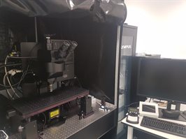
The Intravital Imaging Suite
The Intravital Imaging Suite is a five-microscope suite designed and equipped for high-end intravital microscopy experiments. The equipment is managed by a dedicated facility manager with significant experience in intravital microscopy. Support is provided both for experimental design and the practical skills to deliver intravital microscopy experiments.
IVM-1 Olympus BX61WI - Upright Widefield Microscope
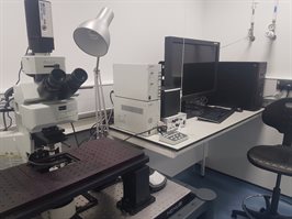
IVM1 is an Olympus BX61 upright widefield microscope that is ideally suited for imaging of transparent tissues such as the cremaster. The microscope has some fluorescent capabilities including an external light source and filters for FITC, CY3 and CY5. The microscope uses Olympus objectives, of which we have x10, x20, x40 and x60 in the facility. There are two detectors attached to the microscope, the Olympus XM10 and Retiga 2000R. The Retiga camera is high quality with a large imaging area, while the Olympus XM10 trades some quality for higher speed. The microscope has a Prior electronic stage, allowing for movement to be controlled by a joystick next to the microscope.
IVM2 - Olympus BX-61WI –Upright Confocal Microscope
IVM-2 is an Olympus BX-61WI upright confocal microscope that is suited to imaging of both transparent tissues and solid tissues which can be positioned flatly for imaging. The confocal nature of the microscope allows for improved imaging in the Z-axis which may be useful for some tissues where the structures of interest are not superficial (if structures are very deep, the multiphoton may be better suited). The microscope is equipped with a Nipkow confocal spinning disk, allowing for real time confocal imaging in live animals. The confocal head is supplied with excitation light from a 3i LaserStack (488nm, 568nm and 647nm lines).
A Photometrics Evolve camera is equipped for image capture as well as a QImaging OptiMOS camera . In addition, this microscope is fitted with a MicroPoint laser ablation system to generate laser damage to microvessels, allowing for the monitoring of thrombus formation during normal tissue perfusion. A warming system is provided for the supply of superfusate buffer at physiological temperature.
IVM-4 - Zeiss Examiner X1 – Upright Confocal Microscope
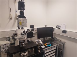
The Zeiss Examiner X1 is an upright confocal microscope that is suited to imaging of both transparent tissues and solid tissues that can be positioned flatly for imaging. The confocal nature of the microscope allows for improved imaging in the Z-axis which may be useful for some tissues where the structures of interest are not superficial (if structures are very deep, the multiphoton may be better suited). The microscope is equipped with a CSU-X1 Nipkow confocal spinning disk head, allowing for realtime confocal imaging in live animals. Compared to the Olympus BX61, the confocal setup on the Zeiss microscope allows the use of lower exposure times than our other microscopes.
The confocal head is supplied by excitation light from a 3i LaserStack (488nm, 568nm, 647nm lines). The confocal head is fitted with a Photometrics Prime BSI detector. The microscope also has a widefield camera and light source, allowing for non-confocal imaging. The microscope also has a laser ablation system attached (Ablate!) for the generation of vascular thrombosis. A warming system is provided with digital temperature feedback for maintaining superfusate buffer at pre-defined temperature. The microscope has a Prior electronic stage, allowing for movement to be controlled by a joystick next to the microscope.
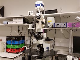
Olympus FLUOView FVMPE-RS – Upright multiphoton Microscope
The FluoView FVMPE-RS is an upright multiphoton microscope. It is a dual-laser system, with a 1040nm fixed beam and a 690nm-1300nm tunable beam. The microscope is also fitted with a separate external stimulation source for optogenetics and photoactivation. These can be used simultaneously, providing true simultaneous excitation of multiple flourophores. The scan head has both Resonant and Galvano scanning modes allowing for the capture of high-quality or high-speed imaging. Identification of localised areas of interest allow scan rates up to 430fps in Resonance mode. There are four detectors on the setup, including two gallium arsenide phosphide (GaAsP) detectors which are considerably more sensitive than conventional PMTs.
The setup has five separate emission filters allowing for the capture of a diverse range of fluorophores. A dichroic mirror is available to split the returning emission light, allowing for the capture of up to four fluorophores simultaneously. The microscope has several objectives available, including a 25x multiphoton-optimised objective which is customised to the multiphoton set up. Smart alignment and digitally controlled internal optics ensures that the entire lightpath is optimised for the wavelength and objectives used. The microscope has a Prior electronic stage, allowing for movement to be controlled by a joystick next to the microscope. This microscope is available for use in non-animal experiments, please contact us for further information.
Olympus IX81 – Inverted Confocal Microscope
The IX81 is an inverted confocal microscope that is best suited to solid tissues which can form a flat surface for imaging from below (either naturally or with manipulation). The microscope has illumination from both a widefield source and a 3i LaserStack (488nm, 568nm, 647nm). The microscope is fitted with a Nipkow spinning disk confocal head which supplies return light to a QImaging OptiMOS camera. As a result of the orientation of this microscope and the sample, all of the objectives available for this setup are air objectives.
Moor FLPI-2 – Full-field laser perfusion imager

The FLPI-2 is a full field laser perfusion imager which utilises Laser Speckle Contrast Imaging to non-invasively monitor tissue perfusion. The device delivers real-time, high-resolution perfusion images that can be obtained from any exposed tissue with minimal additional surgical intervention required. It has three measurement modes with flexible sampling rates (spatial, temporal and sliding window algorithms) to optimise your data for frame rate, spatial resolution and file size. The setup can provide spatial resolution of 10 microns per pixel.
General facilities
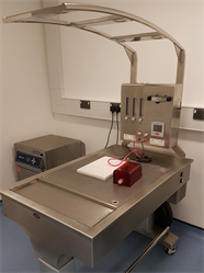
All rooms are equipped with LEEC downdraft tables and VetTech oxygen generators. Each downdraft table has an attached vaporiser and gaseous anaesthetic can be used in most setups if required (please enquire first). The facility has its own dedicated dry heat sterilisation oven which can be used to sterilise tools within the facility.
Training is provided by a dedicated facility manager and NACWO who has significant experience in intravital imaging. All of the machines in the facility are connected to the Research Data Network, allowing fast data transfer between machines and the wider campus network.
Recent publications
Recent publications
Kavanagh D, Gallagher MT, Kalia N. Tify: a quality-based frame selection tool for improving the output of unstable biomedical imaging. PLOS One. 2019 – in press
Noy PJ, Gavin RL, Colombo D, et al. Tspan18 is a novel regulator of the Ca2+ channel Orai1 and von Willebrand factor release in endothelial cells. Haematologica; 2018 – ePub ahead of print
Smith CW, Raslan Z, Parfitt L et al. TREM-like transcript 1: a more sensitive marker of platelet activation than P-selectin in humans and mice. Blood Adv. 2018 Aug 28;2(16):2072-2078.
Sheriff L, Alanazi A, Ward LSC, et al. Origin‐Specific Adhesive Interactions of Mesenchymal Stem Cells with Platelets Influence Their Behavior After Infusion. Stem Cells; 2018 Jul; 36(7): 1062–1074
Mori J, Nagy Z, Di Nunzio G et al. Maintenance of murine platelet homeostasis by the kinase Csk and phosphatase CD148. Blood. 2018 Mar 8;131(10):1122-1144.
Smith CW, Thomas SG, Raslan Z et al. Mice Lacking the Inhibitory Collagen Receptor LAIR-1 Exhibit a Mild Thrombocytosis and Hyperactive Platelets. Arterioscler Thromb Vasc Biol. 2017 May;37(5):823-835
Payne H, Ponomaryov T, Watson SP, et al. Mice with a deficiency in CLEC-2 are protected against deep vein thrombosis. Blood. 2017 Apr 6; 129(14): 2013–2020
King A, Houlihan DD, Kavanagh D, et al. Sphingosine-1-Phosphate Prevents Egress of Hematopoietic Stem Cells From Liver to Reduce Fibrosis. Gastroenterology. 2017 Jul;153(1):233-248.e16.
Yemm A, Adams D, Kalia N. Targeting the delivery of systemically administered haematopoietic stem/progenitor cells to the inflamed colon using hydrogen peroxide and platelet microparticle pre-treatment strategies. Stem Cell Res. 2015 Nov;15(3):569-580. doi: 10.1016/j.scr.2015.10.001. Epub 2015 Oct 9.
Kavanagh D, Suresh S, Newsome PN et al. Pretreatment of Mesenchymal Stem Cells Manipulates Their Vasculoprotective Potential While Not Altering Their Homing Within the Injured Gut. Stem Cells. 2015 Sep;33(9):2785-97
Chimen M, McGettrick HM, Apta B. Homeostatic regulation of T cell trafficking by a B cell-derived peptide is impaired in autoimmune and chronic inflammatory disease. Nat Med. 2015 May;21(5):467-475.
Contact us
Contact us
Contact us via email at: